Showing posts with label Arginine. Show all posts

Sunday, September 23, 2012

Targeted delivery of therapeutics to bone and connective tissues: current status and challenges- Part I

Reporter Aviral Vatsa, PhD MBBS

Annual treatment costs for musculoskeletal diseases in the US are roughly 7.7% (~ $849 billion) of total gross domestic product. Such disorders are the main cause of physical disability in US. Almost half of all chronic conditions in people can be attributed to bone and joint disorders. In addition there is increasing ageing population and associated increases in osteoporosis and other diseases, rising incidences of degenerative intervertebral disk diseases and numbers of revision orthopedic arthroplasty surgeries, and increases in spinal fusions. All these factors contribute towards the increasing requirement of bone regeneration and reconstruction methods and products. Delivery of therapeutic grade products to bone has various challenges. Parenteral administration limits the efficient delivery of drugs to the required site of injury and local delivery methods are often expensive and invasive. The theme issue of Advance Drug Delivery reviews focuses on the current status of drug delivery to bone and the issues facing this field. Here is the first part of these reviews and research articles.

1. Demineralized bone matrix in bone repair: History and use

Abstract

Demineralized bone matrix (DBM) is an osteoconductive and osteoinductive commercial biomaterial and approved medical device used in bone defects with a long track record of clinical use in diverse forms. True to its name and as an acid-extracted organic matrix from human bone sources, DBM retains much of the proteinaceous components native to bone, with small amounts of calcium-based solids, inorganic phosphates and some trace cell debris. Many of DBM's proteinaceous components (e.g., growth factors) are known to be potent osteogenic agents. Commercially sourced as putty, paste, sheets and flexible pieces, DBM provides a degradable matrix facilitating endogenous release of these compounds to the bone wound sites where it is surgically placed to fill bone defects, inducing new bone formation and accelerating healing. Given DBM's long clinical track record and commercial accessibility in standard forms and sources, opportunities to further develop and validate DBM as a versatile bone biomaterial in orthopedic repair and regenerative medicine contexts are attractive.

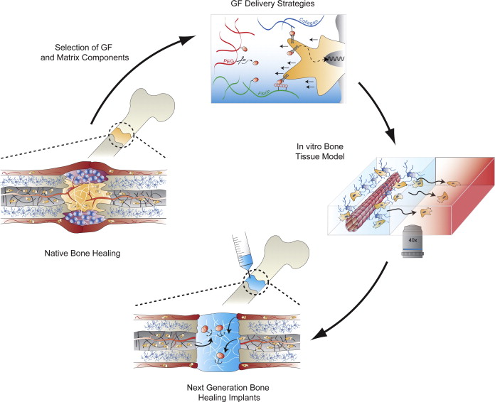
2. Biomimetic hydrogels for controlled biomolecule delivery to augment bone regeneration

Abstract

The regeneration of large bone defects caused by trauma or disease remains a significant clinical problem. Although osteoinductive growth factors such as bone morphogenetic proteins have entered clinics, transplantation of autologous bone remains the gold standard to treat bone defects. The effective treatment of bone defects by protein therapeutics in humans requires quantities that exceed the physiological doses by several orders of magnitude. This not only results in very high treatment costs but also bears considerable risks for adverse side effects. These issues have motivated the development of biomaterials technologies allowing to better control biomolecule delivery from the solid phase. Here we review recent approaches to immobilize biomolecules by affinity binding or by covalent grafting to biomaterial matrices. We focus on biomaterials concepts that are inspired by extracellular matrix (ECM) biology and in particular the dynamic interaction of growth factors with the ECM. We highlight the value of synthetic, ECM-mimicking matrices for future technologies to study bone biology and develop the next generation of ‘smart’ implants.
 

3. Calcium phosphate cements as drug delivery materials

Abstract

Calcium phosphate cements are used as synthetic bone grafts, with several advantages, such as their osteoconductivity and injectability. Moreover, their low-temperature setting reaction and intrinsic porosity allow for the incorporation of drugs and active principles in the material. It is the aim of the present work to: a) provide an overview of the different approaches taken in the application of calcium phosphate cements for drug delivery in the skeletal system, and b) identify the most significant achievements. The drugs or active principles associated to calcium phosphate cements are classified in three groups, i) low molecular weight drugs; ii) high molecular weight biomolecules; and iii) ions.


4. Silk constructs for delivery of musculoskeletal therapeutics

Abstract

Silk fibroin (SF) is a biopolymer with distinguishing features from many other bio- as well as synthetic polymers. From a biomechanical and drug delivery perspective, SF combines remarkable versatility for scaffolding (solid implants, hydrogels, threads, solutions), with advanced mechanical properties and good stabilization and controlled delivery of entrapped protein and small molecule drugs, respectively. It is this combination of mechanical and pharmaceutical features which renders SF so exciting for biomedical applications. This pattern along with the versatility of this biopolymer has been translated into progress for musculoskeletal applications. We review the use and potential of silk fibroin for systemic and localized delivery of therapeutics in diseases affecting the musculoskeletal system. We also present future directions for this biopolymer as well as the necessary research and development steps for their achievement.
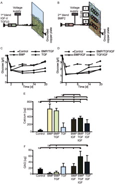
5. Demineralized bone matrix as a vehicle for delivering endogenous and exogenous therapeutics in bone repair

Abstract

As a unique human bone extract approved for implant use, demineralized bone matrix (DBM) retains substantial amounts of endogenous osteoconductive and osteoinductive proteins. Commercial preparations of DBM represent a clinically accessible, familiar, widely used and degradable bone-filling device, available in composite solid, strip/piece, and semi-solid paste forms. Surgically placed and/or injected, DBM releases its constituent compounds to bone sites with some evidence for inducing new bone formation and accelerating healing. Significantly, DBM also has preclinical history as a drug carrier by direct loading and delivery of several important classes of therapeutics. Exogenous bioactive agents, including small molecule drugs, protein and peptide drugs, nucleic acid drugs and transgenes and therapeutic cells have been formulated within DBM and released to bone sites to enhance DBM's intrinsic biological activity. Local release of these agents from DBM directly to surgical sites in bone provides improved control of dosing and targeting of both endogenous and exogenous bioactivity in the context of bone healing using a clinically familiar product. Given DBM's long clinical track record and commercial accessibility in standard forms and sources, opportunities to formulate DBM as a versatile matrix to deliver therapeutic agents locally to bone sites in orthopedic repair and regenerative medicine contexts are attractive.


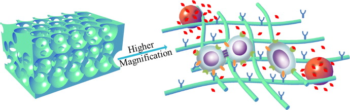
6. Nanofiber-based delivery of bioactive agents and stem cells to bone sites

Abstract

Biodegradable nanofibers are important scaffolding materials for bone regeneration. In this paper, the basic concepts and recent advances of self-assembly, electrospinning and thermally induced phase separation techniques that are widely used to generate nanofibrous scaffolds are reviewed. In addition, surface functionalization and bioactive factor delivery within these nanofibrous scaffolds to enhance bone regeneration are also discussed. Moreover, recent progresses in applying these nanofiber-based scaffolds to deliver stem cells for bone regeneration are presented. Along with the significant advances, challenges and obstacles in the field as well as the future perspective are discussed.


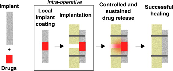
7. Intra-operatively customized implant coating strategies for local and controlled drug delivery to bone

Abstract

Bone is one of the few tissues in the human body with high endogenous healing capacity. However, failure of the healing process presents a significant clinical challenge; it is a tremendous burden for the individual and has related health and economic consequences. To overcome such healing deficits, various concepts for a local drug delivery to bone have been developed during the last decades. However, in many cases these concepts do not meet the specific requirements of either surgeons who must use these strategies or individual patients who might benefit from them. We describe currently available methods for local drug delivery and their limitations in therapy. Various solutions for drug delivery to bone focusing on clinical applications and intra-operative constraints are discussed and drug delivery by implant coating is highlighted. Finally, a new set of design and performance requirements for intra-operatively customized implant coatings for controlled drug delivery is proposed. In the future, these requirements may improve approaches for local and intra-operative treatment of patients.


8. Local delivery of small and large biomolecules in craniomaxillofacial bone
Abstract

Current state of the art reconstruction of bony defects in the craniomaxillofacial (CMF) area involves transplantation of autogenous or allogenous bone grafts. However, the inherent drawbacks of this approach strongly urge clinicians and researchers to explore alternative treatment options. Currently, a wide interest exists in local delivery of biomolecules from synthetic biomaterials for CMF bone regeneration, in which small biomolecules are rapidly emerging in recent years as an interesting adjunct for upgrading the clinical treatment of CMF bone regeneration under compromised healing conditions. This review highlights recent advances in the local delivery small and large biomolecules for the clinical treatment of CMF bone defects. Further, it provides a perspective on the efficacy of biomolecule delivery in CMF bone regeneration by reviewing presently available reports of pre-clinical studies using various animal models.

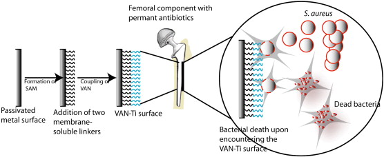
9. Immobilized antibiotics to prevent orthopaedic implant infections

Abstract

Many surgical procedures require the placement of an inert or tissue-derived implant deep within the body cavity. While the majority of these implants do not become colonized by bacteria, a small percentage develops a biofilm layer that harbors invasive microorganisms. In orthopaedic surgery, unresolved periprosthetic infections can lead to implant loosening, arthrodeses, amputations and sometimes death. The focus of this review is to describe development of an implant in which an antibiotic tethered to the metal surface is used to prevent bacterial colonization and biofilm formation. Building on well-established chemical syntheses, studies show that antibiotics can be linked to titanium through a self-assembled monolayer of siloxy amines. The stable metal–antibiotic construct resists bacterial colonization and biofilm formation while remaining amenable to osteoblastic cell adhesion and maturation. In an animal model, the antibiotic modified implant resists challenges by bacteria that are commonly present in periprosthetic infections. While the long-term efficacy and stability is still to be established, ongoing studies support the view that this novel type of bioactive surface has a real potential to mitigate or prevent the devastating consequences of orthopaedic infection.
10. Local delivery of nitric oxide: Targeted delivery of therapeutics to bone and connective tissues

Abstract

Non-invasive treatment of injuries and disorders affecting bone and connective tissue remains a significant challenge facing the medical community. A treatment route that has recently been proposed is nitric oxide (NO) therapy. Nitric oxide plays several important roles in physiology with many conditions lacking adequate levels of NO. As NO is a radical, localized delivery via NO donors is essential to promoting biological activity. Herein, we review current literature related to therapeutic NO delivery in the treatment of bone, skin and tendon repair.


Bibliography

  1. Demineralized bone matrix in bone repair: History and use

  2. Biomimetic hydrogels for controlled biomolecule delivery to augment bone regeneration

  3. Calcium phosphate cements as drug delivery materials

  4. Silk constructs for delivery of musculoskeletal therapeutics

  5. Demineralized bone matrix as a vehicle for delivering endogenous and exogenous therapeutics in bone repair

  6. Nanofiber-based delivery of bioactive agents and stem cells to bone sites

  7. Intra-operatively customized implant coating strategies for local and controlled drug delivery to bone

  8. Immobilized antibiotics to prevent orthopaedic implant infections

  9. Local delivery of nitric oxide: Targeted delivery of therapeutics to bone and connective tissues

Wednesday, September 19, 2012

Discovery of nitric oxide and its role in vascular biology

Curated/reported by : Aviral Vatsa PhD, MBBS

Based on : S Moncada et al

It was in 1980 that Furchgott & Zawadzki first described endothelium- dependent relaxation of the blood vessels by acetylcholine. Further studies in 1984 revealed that other factors such as bradykinin, histamine and 5-hydroxytryptamine release endothelium derived relaxing factor (EDRF), which can modulate vessel tone. EDRF was shown to stimulate soluble guanylate cyclase and was inhibited by haemoglobin. In 1986 it was demonstrated that superoxide (O2-) anions mediated EDRF inactivation and that the inhibitors of EDRF generated superoxide (O2-) anions in solution as a mean to inhibit EDRF. It was later established that all compounds that inhibit EDRF have one property in common, redox activity, which accounted for their inhibitory action on EDRF. One exception was haemoglobin, which inactivates EDRF by binding to it. In 1987 Furchgott proposed that EDRF might be nitric oxide (NO) based on a study of the transient relaxations of endothelium-denuded rings of rabbit aorta to ‘acidified’ inorganic nitrite (NO-) solutions and the observations that superoxide dismutase (SOD, which removes O2-) protected EDRF. Till then NO was not known to be produced in mammalian cells. In 1988 Palmer et al could detect NO production both biologically and chemically by chemiluminescence. The following year in 1989 the enzyme responsible for NO production, NO synthase, was discovered and L-arginine:NO pathway was proposed.

Roles of L-arginine:NO pathway :

By 1987 it was proposed that NO is generated in tissues other than endothelium. Hibs et al and Marletta et al proposed that NO was generated by macrophages. Moreover release of EDRF was demonstrated in cerebellar cells following activation with N-methyl-D- aspartate (NMDA ). Both noradrenergic and cholinergic responses are ‘controlled’ by the nitrergic system so that the release of NO (e.g., during electrical field stimulation) counteracts and dominates the response to the noradrenergic or cholinergic stimulus (Cellek & Moncada, 1997). Mechanism of penile erection was unveiled by the studies on nitrergic neurotransmission that led to therapeutic intervention. Selective damage of nitrergic nerves in disease states was proposed as a potent mechanism of pathophysiology. Broadly three areas of research based on three isoforms of NOS came into being; cardiovascular, nervous and immunology. Identification of NG-monomethyl-L-arginine (L-NMMA) as an inhibitor of the synthesis of NO lay the basis of future research into investigating the role of NO in biological systems. In 1989 it was demonstrated that intravenous infusion of L-NMMA resulted in increase in blood pressure that was reversible by infusing L-arginine. NO was thus implicated in constantly maintaining blood vessel tone. eNOS knockout studies showed a hypertensive phenotypes in the animal models and over expression of eNOS led to lowering of the blood pressure. Furthermore, eNOS activation was attributed to phosphorylation of a specific tyrosine residue in the enzyme.

NO and mitochondria: (detailed here)

NO reacts with some of the complexes of the respiratory chain, and inhibits mitochondrial respiration- this is a well accepted notion. Initially it was believed that the target for NO was soluble guanylate cyclase, which in vasculature would lead to elevation of cGMP that eventually results in NO mediated vasodilatation and platelet aggregation inhibition. In 1994, another potential target, cytochrome c oxidase, for inhibitory effects of NO was discovered. This was a reversible effect, in competition with oxygen concentrations. Increases in NO production were also shown to inhibit cellular respiration irreversibly by selectively inhibiting complex I . Hence in 2002 it was proposed that this might be a mechanism through which cell pathology was initiated in certain conditions. Furthermore, NO was proposed to be implicated in the activation of the grp78-dependent stress response , via modulating calcium -related interaction between mitochondria and endoplasmic reticulum . This host defence mechanism might also have role in vasculature. Further evidence was provided in 2003 to link the role of NO in mitochondrogenesis and thus indicating that NO might be involved in the regulation of the balance between glycolysis and oxidative phosphorylation in cells.

NO and pathophysiology :

Lack of NO: By 2000, NO was established as a haemostatic regulator in the vasculature. Its absence was implicated in pathological states such as hypertension and vasospasm. These pathophysiological states share a common beginning of endothelial dysfunction, which has low NO production as one of its characterstic features. This dysfunction has been observed prior to the appearance of cardiovascular disease in predisposed subjects with family history of essential hypertension and atherosclerosis. The most likely mechanism for endothelial dysfunction is that of a reduced bioavailability of NO . The mechanism of this aspect is discussed elsewhere on this site. Protection against reduction of NO bio-availability in the vasculature is a vital therapeutic target and is extensively explored. This can be achieved by the use of antioxidants and/or augmentation of eNOS expression. In 2003 statins were shown to increase the production of endothelial NO in endothelial cell cultures and in animals by the reduction of oxidative stress or by increasing the coupling of the eNOS. It was way back in 1994 that oestrogen was shown to increase both the activity and expression of eNOS. In addition, more recently in 2003, oestrogen was shown to reduce the breakdown of available NO.

Excess of NO: In 2000 it was shown that NO produced from iNOS in vasculature is involved in extensive vasodilatation in septic shock. Later it was demonstrated that inhibition of mitochondrial respiration is an important component of the NO-induced tissue damage. This inhibition of respiration, which is initially NO-dependent and reversible, becomes persistent with time as a result of oxidative stress . Such metabolic hypoxic states where in tissues cannot utilise available oxygen due to NO, could also could also contribute to other inflammatory and degenerative conditions. An obvious therapeutic target for reducing NO production in such conditions would be L-NMMA. L-NMM was tested in a clinical trial for septic shock in 2004. The results were however disappointing probably due to the blanket reduction in NO production from other NOS enzymes there by having deleterious effects on the treatment group. More specific inhibitors for NOS forms are being investigated for in different disease states.

In conclusion, the L-arginine: NO pathway has had a major impact in many areas of research, specially vascular biology. A lot has been understood about this pathway and its interactions, therapeutic targets are being aggressively investigated, but further investigations are required to delineate further the role of NO in human health and disease.

Further reading

http://www.ncbi.nlm.nih.gov/pmc/articles/PMC1760731/?tool=pubmed

Nitric Oxide and Platelet Aggregation

Inhaled NO in Pulmonary Artery Hypertension and Right Sided Heart Failure

Cardiovascular Disease (CVD) and the Role of agent alternatives in endothelial Nitric Oxide Synthase (eNOS) Activation and Nitric Oxide Production

Nitric Oxide in bone metabolism

Nitric oxide and signalling pathways

Rationale of NO use in hypertension and heart failure

Interaction of Nitric Oxide and Prostacyclin in Vascular Endothelium

Nitric Oxide has a ubiquitous role in the regulation of glycolysis -with a concomitant influence on mitochondrial function

Tuesday, September 11, 2012

Nitric oxide: role in Cardiovascular health and disease

Authour/Curator: Aviral Vatsa PhD MBBS

This post is in continuation with posts on basics of NO metabolism and its effect on physiology. Other topics are covered under the following posts.

  1. Nitric Oxide and Platelet Aggregation

  2. Inhaled NO in Pulmonary Artery Hypertension and Right Sided Heart Failure

  3. Cardiovascular Disease (CVD) and the Role of agent alternatives in endothelial Nitric Oxide Synthase (eNOS) Activation and Nitric Oxide Production

  4. Nitric Oxide in bone metabolism

  5. Nitric oxide and signalling pathways

  6. Rationale of NO use in hypertension and heart failure

Nitric oxide plays wide variety of roles in cardiovascular system and acts as a central point for signal transduction pathway in endothelium. NO modulates vascular tone, fibrinolysis, blood pressure and proliferation of vascular smooth muscles. In cardiovascular system disruption of NO pathways or alterations in NO production can result in preponderance to hypertension, hypercholesterolemia, diabetes mellitus, atherosclerosis and thrombosis. The three enzyme isoforms of NO synthase family are responsible for generating NO in different tissues under various circumstances. The endothelial NOS (eNOS) is expressed in endothelial cells, the inducible NOS (iNOS) is expressed in macrophages and neuronal NOS (nNOS) is expressed in certain neurons and skeletal muscle. Although the basic mechanism of action for NO production is the same for all three NOS isoforms, yet deficiencies of each one of them manifest differently or with varying severity in the body e.g. eNOS deficiency might lead to hypertension, more severe form of vascular injury to cerebral ischaemia and more severe form of atherosclerosis induced by hypercholesterolemic diet whereas nNOS deficiency might show less severe form of vascular injury to cerebral ischaemia and absence of iNOS might lead to reduced hypotension in septic shock.

Reduction in NO production is implicated as one of the initial factors in initiating endothelial dysfunction. This reduction could be due to

  • reduction in eNOS production

  • reduction in eNOS enzymatic activity

  • reduced bioavailability of NO

eNOS production is increased by physiological sheer stress on endothelial cells resulting from normal flow of blood along the arterial walls. Alterations in fluid sheer stress patterns e.g due to arterial constriction has been shown to have detrimental effect on eNOS production in endothelial cells. eNOS production is decreased by LDL, angiotensin II and TNF alpha. eNOS is tightly coupled enzyme and its activity can be significantly reduced by reduction in availability of cofactors and substrates, and by competitive inhibitors such as ADMA. Furthermore, uncoupling of eNOS can result in increased production of reactive species of both oxygen (superoxide) and nitrogen (peroxinitrite), which inturn can further reduce eNOS bioavailability. A range of therapeutic targets aim at increasing bioavailability of eNOS and they are summarised here.

Increased production of ROS and peroxinitrite is associated with endothelial dysfunction. Coronary heart disease risk factors may increase NOS mediated ROS formation and peroxinitrite formation. Such risk factors are associated with decreased NO production levels in the vasculature. However, recent data suggests that reduction in bioavailable NO levels in the arteries could be due to increased local oxidative stress rather than reduction in basal NO production.

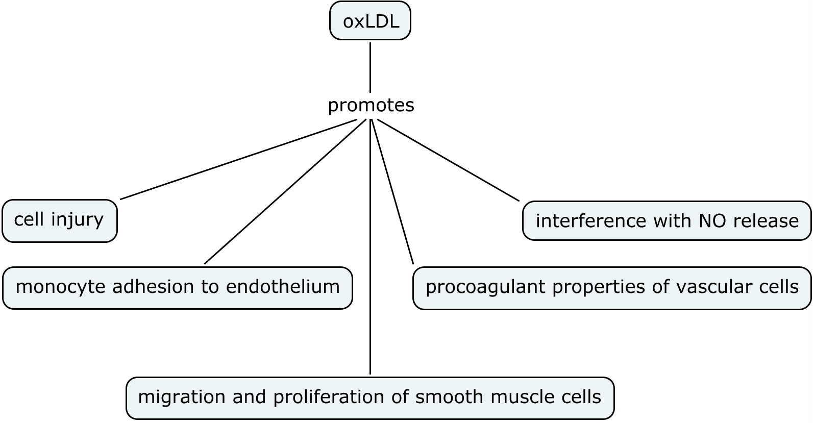
Oxidation dependent mechanisms have been implicated in endothelial dysfunction. Oxidized low density lipo-proteins (oxLDL) play an important role in early endothelial dysfunction and hence early atherosclerosis (see figure below)

oxLDL can uncouple eNOS and reduced uptake of L-ariginine that can lead to production of superoxide radical oxygen. OxLDL can interfere with NO production and lead to altered NO signalling in the vascular endothelium. In addition, different arteries can be affected differently by these physiological changes e.g. oxLDL affects carotid artery and not the basilar artery thereby implying that intracranial arteries might be protected from endothelium-mediated oxidative injury and hence atherosclerosis. And finally NO can modulate oxidation mediated apoptotic signals in the vessel wall. Hence atherosclerosis can result from the derangement of fine imbalance between NO bioavailability and local oxidative stress.

Therapeutic targets:

There are various pathways being targeted to modulate the bioavailability of eNOS and NO such as

  1. Recoupling of eNOS to cofactors and substrates

  2. Modulation of eNOS activity by genomic and non genomic mechanisms e.g. by statins, ACE inhibitors, angiotensin II receptor blockers, calcium channel blockers , KLF2 modulators

  3. The suppression of inflammatory signalling pathways by PPAR-α activation

  4. Modulation of caeviolin mediated endocytosis and thus dissociation of eNOS from caevolin

The above list is not exhaustive, but this post here summarises recent developments in therapeutic targets in NO /eNOS regulation.

Based on:

Nitric Oxide and Pathogenic Mechanisms Involved in the Development of Vascular Diseases

Claudio Napoli and Louis J. Ignarro

Arch Pharm Res Vol 32, No 8, 1103-1108, 2009 , DOI 10.1007/s12272-009-1801-1 


Thursday, August 23, 2012

Nitric Oxide Signalling Pathways

Curator/ Author: Aviral Vatsa, PhD, MBBS

In continuation with the previous posts that dealt with short history and chemistry of nitric oxide (NO), here I will try to highlight the pathways involved in NO chemical signalling.

NO is a very small molecule, with a short half life (<5 sec). It diffuses rapidly to its surroundings and is metabolised to nitrites and nitrates. It can travel short distances, a few micrometers, before it is oxidised. Although it was previously believed that NO can only exert its effect for a very short time as other nitrogen oxides were believed to be biologically inert. Recent data suggests that other NO containing compounds such as S- or N-nitrosoproteins and iron-nitrosyl complexes can be reduced back to produce NO. These NO containing compounds can serve as storage and can reach distant tissues via blood circulation, remote from their place of origin. Hence NO can have both paracrine and 'endocrine' effects.

Intracellularly the oxidants present in the cytosol determine the amount of bioacitivity that NO performs. NO can travel roughly 100 microns from NOS enzymes where it is produced. NOS enzymes on the other hand are localised to specific sub-cellular areas, which have relevant proteins in the vicinity as targets for signalling.

NO signalling occurs primarily via three mechanisms (according to Martínez-Ruiz et al):

  1. Classical: This occurs via soluble guanylyl cyclase (sGC). Once NO is produced by NOS it diffuses to sGC intracellularly or even in other cells. SGC is highly sensitive for NO, even nanomolar amounts of NO activates sGC, thus making it a potent target for NO in signalling pathways. sGC in turn increases the conversion of GTP to cGMP. cGMP further mediates the regulation of contractile proteins and gene expression pathways via cGMP-activated protein kinases (PKGs). cGMPs cause confirmational changes in PKGs. Signalling by cGMP is terminated by the action of phosphodiestrases (PDEs). PDEs have become major therapeutic targets in the upcoming exciting research projects.
  2. Less classical: Within the mitochondria NO can compete with O2 and inhibit cytochrome c oxidase (CcO) enzyme. This is a reversible inhibition that depends on O2and NO concentrations and can occur at physiological levels of NO. Various studies have demonstrated that endogenously generated NO can inhibit respiration or that NOS inhibitors can increase respiration at cellular, tissue or whole animal level. Although the exact mechanism of CcO inhibition of NO is still debated, NO-CcO interaction is considered important signalling step in a variety of functions such as inhibition of mitochondrial oxidative phosphorylation, apoptosis and reactive oxygen species (ROS) generation. Interestingly, at higher concentration (~1nM) NO can cause irreversible inhibition of cellular oxidation by reversible and/or irreversible damage to the mitochondrial iron–sulfur centers,In addition to the above mentioned pathways, NO (along with AMP, ROS and O2), can also activate AMP- activated protein kinase (AMPK), an enzyme that plays a central role in regulating intracellular energy metabolism. NO can also regulate hypoxia inducible factor (HIF), an O2-dependent transcription factor that plays a key role in cell adaptation to hypoxia .
  3. Non- classical: S-nitrosylation or S-nitrosation is the covalent insertion of NO into thiol groups such as of cysteine residues of proteins. It is precise, reversible, and spatiotemporally restricted post translational modification. This chemical activity is dependent upon the reactivity between nitrosylating agent (a small molecule) and the target (protein residue). It might appear that this generic interaction results in non-specific, wide spread chemical activity with various proteins. However, three factors might determine the regulation of specificity of s-nitrosylation for signalling purposes:
  • Subcellular compartmentalisation: high concentrations of nitrosylating agents are required in the vicinity of target residues, thus making it a specific activity.
  • Site specificity: certain cysteine residues are more reactive in specific protein microenvironments than others, thus favouring their modification. As a result under physiological conditions only a specific number of cysteine residues would be modified, but under higher NO levels even the slow reacting ones would be modified. Increased impetus in research in this area to determine protein specificity to s-nitrosylation provides huge potential in discovering new therapeutic targets.
  • Denitrosylation: different rates of denitrosylation result in s-nitrosylation specificity.

Other modifications in non classical NO mechanisms include S-glutathionylation and tyrosine nitration

Peroxynitrite: It is one of the important reactive nitrogen species that has immense biological relevance. NO reacts with superoxide to form peroxynitrite. Production of peroxynitrite depletes the bioactivty of NO in physiological systems. Peroxynitrite can diffuse through membranes and react with cellular components such as mitochondrial proteins, DNA, lipids, thiols, and amino acid residues. Peroxynitrite can modify proteins such as haemoglobin, myoglobin and cytochrome c. it can alter calcium homeostasis and promote mitochondrial signalling of cell death. However, NO itself in low concentrations have protective action on mitochondrial signalling of cell death.

More details about various aspects of NO signalling can be obtained from the following references.

The post is based on the following Sources:

  1. http://www.sciencedirect.com/science/article/pii/S089158491100236X

    http://dx.doi.org/10.1016/j.freeradbiomed.2011.04.010

  2. http://content.karger.com/produktedb/produkte.asp?doi=338150

    Cardiology 2012;122:55-68 (DOI: 10.1159/000338150)

  3. http://content.onlinejacc.org/article.aspx?articleid=1137266

     J Am Coll Cardiol. 2006;47(3):580-581. doi:10.1016/j.jacc.2005.11.016

  4. http://goo.gl/y6oY3


In addition, other aspects of NO involvement in biological systems in humans are covered in the following posts on this site:

  1. Nitric Oxide and Platelet Aggregation
  2. Inhaled NO in Pulmonary Artery Hypertension and Right Sided Heart Failure
  3. Cardiovascular Disease (CVD) and the Role of agent alternatives in endothelial Nitric Oxide Synthase (eNOS) Activation and Nitric Oxide Production
  4. Nitric Oxide in bone metabolism

Wednesday, July 25, 2012

Nitric Oxide production in Systemic sclerosis


Curator: Aviral Vatsa, PhD, MBBS
Systemic sclerosis (SSc) is a type of autoimmune disease when the body’s immune system attacks and destroys body’s healthy tissue. It is characterised by lesions in the vessels and accumulation of collagen in the tissues. Although the pathogenesis of this disease is not clear, but one of the suggestions is that the endothelium fails to produce NO upon cold stimulation. Physiologically, NO acts as a vasodilator and its deficiency has been implicated in diseases such as hypertension and atherosclerosis.
In the body NO is generated when L-arginine is converted to L-citruline in the presence of NO synthase (NOS) enzyme, molecular oxygen, NADPH, and other cofactors. Principally, three isoenzymes of NOS are present in the body to catalyse the production of NO in various anatomic locations and under various physiological conditions. Three distinct genes encode for the three types of NOS i.e. endothelial (eNOS or NOS-3), neuronal (nNOS or NOS-1), and inducible (iNOS or NOS-2) NOS.
The inducible type 2 NOS (iNOS) may act as an immunoregulator. Several reports have provided evidence for the existence of a NO pathway in human mononuclear cells.
It is not well established if NO production increases or decreases in SSc patients. In one study by Allanore et al, NO production was shown to be reduced in plasma and PBMC supernatants, and iNOS synthesis in PBMCs.
The authors of this study investigated NO metabolites in plasma and PBMC supernatants, and iNOS synthesis in PBMC to see if the level of NO production by peripheral blood mononuclear cells (PBMC) was low in SSc, as this might contribute to the vasodilatory abnormalities observed in this disease.
Eighteen patients with SSc were compared with two control groups: 16 patients with rheumatoid arthritis (RA) and 23 patients with mechanical sciatica.The NO metabolites nitrite and nitrates were determined by flurometeric and spectrometeric assays respectively. iNOS expression was determined by using monoclonal anti‐NOS2 antibody and FACS analyses.
The data suggested a decrease in plasma NO concentration and iNOS production in PBMC in patients with systemic sclerosis as compared with patients with rheumatoid arthritis and sciatica. Subgroup analysis showed no difference between limited and diffuse SSc forms. Total plasma nitrite concentrations in five healthy volunteers were similar to those in patients with sciatica, which is consistent with this group being an appropriate control group.
Thus the authors suggest that low NO production in Ssc patients might be involved in the tendency towards vasospam.
However in other studies authors have shown an increase in NO production in SSc patients. Takagi et al set out to investigate this discrepancy in NO production in SSc patients. They sought to determine whether increased NO levels are associated with various clinical subsets of SSc patients, and to assess the contribution of fibroblasts in skin lesions to NO synthesis.
In this study Takagi et al measured the levels of serum NO metabolites in SSc patients and determined the contribution of the excessive production of NO synthase (NOS)-2 by skin fibroblasts to NO synthesis. Serum NO levels of 45 patients with SSc were significantly higher than those of 20 healthy volunteers. In addition, some clinical features of SSc (the extent of skin fibrosis, short disease duration, and the complication of active fibrosing alveolitis) were all correlated positively with the levels of NO metabolites in SSc patients. RT PCR was used to determine NOS-2 mRNA expression levels in cultured fibroblasts derived from SSc patients.
The authors showed that serum NO levels were significantly elevated in patients with SSc as compared to healthy normal controls. They also demonstrated that NOS-2 was produced spontaneously by cultured SSc fibroblasts, suggesting that increased serum NO levels might reflect in part the elevated expression of NOS-2 by fibroblasts derived from SSc patients.
The discrepancy in NO production could be explained by disease stage, severity of tissue fibrosis and various circumstances of endothelial damage. Takagi et al found increased NO production in early stages of SSc with tissue fibrosis and not in later stages of the disease. Hence they suggest that NO levels may be a sensitive marker of the early stages of the development of severe tissue fibrosis in SSc patients, although a longitudinal and prospective study is needed to confirm this.
NO is known to have dual functions in the body, both beneficial and cyototoxic. Generally it depends upon the concentration and the duration of NO production. Similarily in SSC, the dual functions of NO seem to be both beneficial (as a vasodilator) and harmful (as a cytotoxic effector) in regard to the clinical manifestations of SSc. One of the limitations of these studies is that they did not investigate the absolute concentrations of NO production but only its metaoblites were measured. This might be due the fact that NO is a short-lived molecule (half-life < 5 s) and it is challenging to quantify NO production at single cell level. Such techniques (e.g.DAR 4M AM flurophore) have been developed but are challenging to apply to various experimental set ups. DAR 4M AM has been used to quantify NO production online in single cells. In SSc determination of absolute NO concentrations at cellular or tissue level at various stages of the disease process will go a long way in solving the discrepancy of NO production in SSc patients.
Sources
Related Posts Plugin for WordPress, Blogger...